DUAL SCAN PROTOCOL
BENEFITS OF DUAL SCAN PROTOCOL
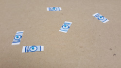
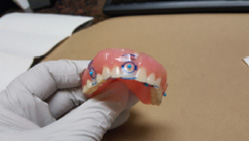
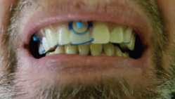
Using CBCT markers eliminates lab costs associated with the need to duplicate patient’s radiopaque acrylic dentures or adding metal bb and hole drilling for gutta-percha.The following instructions ensures surgical guides will mirror the image of the denture andfit the patient’s mouth like the original prosthesis.
Address: 3144 John R Road, Suite 100 Troy, MI 48083A
Phone: 248-740-7770
Toll-Free: 855-361-3335
Email: info@cinzara.com
Web: www.cinzara.com
CBCT IMAGING PROTOCOL FOR PATIENT SCANNING SURGICAL GUIDES
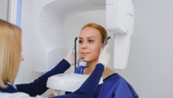
As practitioners work to meet the growing demand for implants, cone beam computed tomography (CBCT) is an essential tool for treatment planning and post-procedure monitoring. By providing highly accurate 3D images of the patient’s anatomy from a single, low-radiation scan, CBCT technology delivers a comprehensive understanding of the patient’s jaw and the anatomical structures necessary to provide proper treatment. The following is a recommended procedure to obtain accurate CBCT scan.
Address: 3144 John R Road, Suite 100 Troy, MI 48083A
Phone: 248-740-7770
Toll-Free: 855-361-3335
Email: info@cinzara.com
Web: www.cinzara.com
SECURING ACCURATE IMPRESSIONS FOR SURGICAL GUIDES
Achieving an accurate and precise guided surgery depends on an accurate impression.
The following factors are important to remember to strategically print accurate surgical guides – regardless of the impression material you use.
PREPPING THE PATIENT
Prepping the Teeth: Ensure the process begins with clean brushed, and flossed teeth.
Before Taking the Impression: Ensure that the teeth are dry free from the presence of blood or saliva and that the tongue and cheeks are distant from the area of interest.
USING INTRAORAL SCAN (HIGHLY RECOMMENDED METHOD)
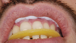
Using Intraoral Scan: As compared to traditional dental impression options, intraoral scan results in surgical guides that fit over the teeth and soft tissue, are more accurate and are distortion-free. (See Figure 1).
STL Files: : After completing the intraoral scans, STL files of the dental arches need to be uploaded to your patient’s file in the secured CinZara account.
USING IMPRESSIONS
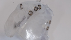
Using PVS Impression Material: Accurate models and impression material help ensure the computer guided dental surgery to go as planned. Therefore, choosing PVS impression material is highly recommended as it reduces the risk of creating an unstable surgical guide. (See Figure 2).
Using Alginate: Taking impressions using alginate poses possible imperfections with the impression, which ultimately affects fabricating an accurate surgical guide. Factors that affect achieving a quality impression include the materials used, the consistency of the mixture, and the experience level of the person mixing the substance to be able to identify the right consistency.
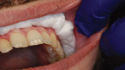
Ensure Accurate Impressions: Ensure the impression is free of any bubbles, include the entire arch and the area of interest on the antagonist and the last tooth near the edentulous area. Using the upper and lower impressions, along with the bite registration, will help us complete the treatment plan using the proper restoration to direct the abutments for final restorations and the impression accurately. (See Figure 3).
The Models: If impressions are used, it is crucial to follow the manufacturer’s instructions to ensure the models are perfect. The use of bubble wrap is important to secure the models and prevent damage during shipment.
Address: 3144 John R Road, Suite 100 Troy, MI 48083A
Phone: 248-740-7770
Toll-Free: 855-361-3335
Email: info@cinzara.com
Web: www.cinzara.com


